- Home

- Reproductive health

- Transvaginal Ultrasound: A Non-Invasive Tool for Early Detection of Reproductive Health Issues
In this Article
Reproductive health
Transvaginal Ultrasound: A Non-Invasive Tool for Early Detection of Reproductive Health Issues
Updated on 9 June 2023


Medically Reviewed by
Dr. Shruti Tanwar
C-section & gynae problems - MBBS| MS (OBS & Gynae)
View Profile

A transvaginal ultrasound is a non-invasive procedure that uses sound waves to examine your reproductive organs from within. This innovative technique has revolutionized the way we detect and diagnose reproductive health issues, allowing healthcare professionals to gather essential information with ease.
Whether you're planning to start a family or simply want to monitor your reproductive well-being, TVS ultrasound is here to empower you with early detection and comprehensive insights. Let's begin our voyage to explore the wonders of transvaginal ultrasound and uncover its potential in safeguarding your reproductive health.
What is a Transvaginal Ultrasound?
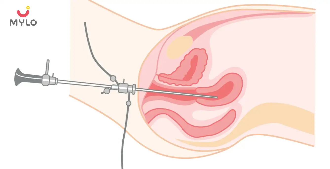
A transvaginal ultrasound is a diagnostic imaging technique that involves the use of a small, probe-like device called a transducer, which is inserted into the vagina. This procedure allows for a closer and more detailed examination of the pelvic organs, including the uterus, ovaries, and fallopian tubes.
By emitting high-frequency sound waves, the transducer creates real-time images of the reproductive organs, providing valuable information about their structure, size, and any potential abnormalities. TVS ultrasound is a safe and effective tool used by healthcare professionals to assess and diagnose various reproductive health conditions.
How is it Different from a Normal Ultrasound?
- A transvaginal ultrasound, also called an endovaginal ultrasound, is a specialized imaging technique used to examine the female reproductive organs. It differs from a normal ultrasound in that it involves the insertion of a transducer into the vagina for a closer view of the pelvic structures.
- The transvaginal approach provides higher-resolution images, allowing for better visualization of the uterus, ovaries, and surrounding tissues. It is particularly useful for assessing conditions such as fibroids, cysts, and abnormalities in the reproductive organs.
- TVS ultrasounds are commonly used during early pregnancy to monitor the development of the fetus and detect any potential complications.
- Many women wonder is TVS ultrasound painful but the procedure is safe, non-invasive, and generally well-tolerated by patients.
What is TVS ultrasound used for?
Transvaginal ultrasound is used for various purposes in the field of reproductive health. Some common uses include:
1. Early Pregnancy Evaluation
TVS is highly effective in confirming early pregnancy, assessing gestational age, and detecting the presence of a fetal heartbeat.
2. Fertility Evaluation
TVS help evaluate the health and condition of the ovaries, uterus, and fallopian tubes, identifying any abnormalities or conditions that may affect fertility.
3. Diagnosis of Gynecological Conditions
TVS aids in diagnosing conditions such as uterine fibroids, ovarian cysts, polycystic ovary syndrome (PCOS), endometriosis, and pelvic inflammatory disease (PID).
4. Monitoring Ovulation
TVS is used to track the development and release of eggs during ovulation, providing valuable information for fertility treatments and assisted reproductive technologies.
5. Evaluation of Abnormal Bleeding
TVS helps assess the thickness and condition of the uterine lining and identify the cause of abnormal uterine bleeding, such as hormonal imbalances or uterine polyps.
6. Guiding Procedures
TVS can guide certain minimally invasive procedures, such as egg retrieval during in vitro fertilization (IVF) or the placement of intrauterine devices (IUDs).
What to Expect During a Transvaginal Ultrasound?
Here's what you can expect during a TVS ultrasound:
1. Preparation
You may be asked to empty your bladder before the procedure to improve the visibility of the pelvic organs. It's also recommended to wear comfortable clothing.
2. Private Setting
You will be taken to a private examination room where the TVS will be performed. The healthcare provider will ensure your privacy and make you feel comfortable throughout the procedure.
3. Positioning
You will be asked to lie down on an examination table and place your feet in stirrups, similar to a gynecological exam. This position allows for easier access to the vagina.
4. Transducer Insertion
A specialized transducer, covered with a disposable protective sheath, will be inserted into the vagina. The transducer emits and receives sound waves to create images of the pelvic organs.
5. Lubrication
To ease the insertion of the transducer, a small amount of gel or lubricant will be applied to the transducer and your vaginal area. This helps ensure smooth movement and comfortable positioning of the transducer.
6. Imaging Process
The transducer will be gently moved within the vagina to obtain images of the uterus, ovaries, and other pelvic structures. You may experience slight pressure or mild discomfort during this process, but it should not be overly painful.
7. Image Interpretation
The healthcare provider will interpret the images in real-time, assessing the condition and health of your reproductive organs. They may point out specific structures or discuss any findings with you.
8. Duration
The entire TVS procedure typically lasts about 15-30 minutes, although this can vary depending on individual circumstances and the specific purpose of the examination.
After the procedure, you can usually resume your daily activities without any restrictions. The healthcare provider will discuss the results with you and may recommend further tests or treatments based on the findings.
Is TVS Ultrasound Painful?
Transvaginal ultrasound (TVS) is generally not considered a painful procedure. However, some women may experience mild discomfort or pressure during the insertion of the transducer into the vagina. The sensation can be likened to that of a gynecological exam or a slight cramping feeling.
It's important to communicate with your healthcare provider throughout the procedure. They can adjust the position and movement of the transducer to minimize any discomfort. If you experience any excessive pain or if the discomfort becomes too intense, it's essential to inform your healthcare provider immediately.
Are there Any Risks of a TVS Ultrasound?
Transvaginal ultrasound (TVS) is considered a safe procedure with minimal risks. However, as with any medical procedure, there are a few potential risks to be aware of:
1. Discomfort or pain
Some women may experience mild discomfort or pressure during the insertion of the transducer. However, this is generally well-tolerated and temporary.
2. Infection
While rare, there is a small risk of infection associated with any invasive procedure. However, healthcare providers take precautions to minimize this risk by using sterile equipment and following proper sterilization procedures.
3. Vaginal bleeding
In rare cases, TVS may cause slight vaginal bleeding. This is usually minimal and resolves on its own. However, if you experience heavy or persistent bleeding, it's important to notify your healthcare provider.
How to Interpret the Results of an Endovaginal Ultrasound?
Interpreting the results of an endovaginal ultrasound requires expertise and knowledge in ultrasound imaging. It is typically performed by a trained healthcare professional, such as a radiologist or gynecologist, who will analyze the images obtained during the procedure. Here are some key points to consider when interpreting the results:
1. Image analysis
The healthcare professional will evaluate the images captured during the ultrasound and examine the structures and organs of interest, such as the uterus, ovaries, and surrounding tissues. They will assess the size, shape, and texture of these structures, looking for any abnormalities or changes.
2. Comparisons with normal findings
The healthcare professional will compare the observed findings with the expected normal anatomy and characteristics. They will consider factors such as age, menstrual cycle phase, and any relevant medical history to make appropriate assessments.
3. Identification of abnormalities
The healthcare professional will look for any signs of abnormalities, such as cysts, tumors, fibroids, or other growths. They will also evaluate the blood flow patterns using Doppler ultrasound, if necessary, to assess the vascularity of certain structures.
4. Clinical correlation
The results of the endovaginal ultrasound will be correlated with your symptoms, medical history, and any other diagnostic tests or imaging studies you may have undergone.
You may like: Pelvic Exam: Details, Outcomes & Dos & Don'ts
Final Thoughts
In conclusion, transvaginal ultrasound is a valuable and non-invasive tool for early detection and evaluation of various reproductive health issues. TVS ultrasound is particularly useful in diagnosing conditions such as ovarian cysts, fibroids, polyps, and assessing fertility-related concerns.
References
1. Kaur, A., & Kaur, A. (2011). Transvaginal ultrasonography in first trimester of pregnancy and its comparison with transabdominal ultrasonography. Journal of Pharmacy and Bioallied Sciences, 3(3), 329.
2. Nahlawi, S., & Gari, N. (2023). Sonography Transvaginal Assessment, Protocols, and Interpretation. PubMed; StatPearls Publishing.


Medically Reviewed by
Dr. Shruti Tanwar
C-section & gynae problems - MBBS| MS (OBS & Gynae)
View Profile


Written by
Madhavi Gupta
Dr. Madhavi Gupta is an accomplished Ayurvedic doctor specializing in Medical content writing with an experience of over 10 years.
Read MoreGet baby's diet chart, and growth tips

Related Articles
How Respiratory Syncytial Virus (RSV) Impacts Premature Babies Differently: What Every Parent Needs To Know
Adverbs: A Comprehensive Guide to help small children learn the usage of adverbs
Expand Your Child's Vocabulary with words that start with X: Easy, Positive, and Engaging Words, Animals, Countries, and Fruits
Unlocking Language Proficiency: The Ultimate Guide to Top 100 Sight Words for Kindergarten and Beyond
Related Questions
Hello frnds..still no pain...doctor said head fix nhi hua hai..bt vagina me pain hai aur back pain bhi... anyone having same issues??

Kon kon c chije aisi hai jo pregnancy mei gas acidity jalan karti hain... Koi btayega plz bcz mujhe aksar khane ke baad hi samagh aata hai ki is chij se gas acidity jalan ho gyi hai. Please share your knowledge

I am 13 week pregnancy. Anyone having Storione-xt tablet. It better to have morning or night ???

Hlo to be moms....i hv a query...in my 9.5 wk i feel body joint pain like in ankle, knee, wrist, shoulder, toes....pain intensity is high...i cnt sleep....what should i do pls help....cn i cosult my doc.

Influenza and boostrix injection kisiko laga hai kya 8 month pregnancy me and q lagta hai ye plz reply me

RECENTLY PUBLISHED ARTICLES
our most recent articles

Diet & Nutrition
New Mom Diet Plan – Month 11 Week 42

Male Infertility
Ejaculatory Duct Obstruction: How It Affects Male Fertility and What You Can Do About It

Testicular Ultrasound: What You Need to Know About the Procedure and Its Benefits

Symptoms of Low AMH to Watch Out For: A Health Alert for Women Trying to Conceive

Medical Procedures
Hysteroscopy: Everything You Need to Know About This Minimally Invasive Procedure

Ayurveda & Homeopathy
Dalchini: How This Herb Can Make Way From Your Spice Rack to Your Medicine Cabinet
- Fenugreek Powder: Health Benefits of Fenugreek From Your Kitchen to Your Medicine Cabinet
- Moringa Powder: The Superfood You Need in Your Diet for a Healthy Lifestyle
- Genital Herpes: Causes, Symptoms, Risks & Treatment
- Ashokarishta: All You Need to Know About This Miracle Tonic for Women
- 10 Amazon Prime Movies to Look Forward to in 2023
- 10 Best Netflix Movies to Watch Out For in 2023
- Fertility Yoga: A Natural Solution to Boost Your Chances of Conception
- How to Get Regular Periods Naturally: Ayurvedic Herbs, Lifestyle Changes & Homeopathy
- Lodhra: The Wonder Herb for Women's Health
- Malabsorption Syndrome: Types, Causes, Symptoms, & Treatment
- Top 10 Short Bedtime Stories for Kids
- RH Incompatibility in Pregnancy - Causes, Symptoms & Treatments
- Patent Ductus Arteriosus (PDA) Symptoms & Treatment
- Why Babies Cry After Birth?


AWARDS AND RECOGNITION
Mylo wins Forbes D2C Disruptor award
Mylo wins The Economic Times Promising Brands 2022
AS SEEN IN


At Mylo, we help young parents raise happy and healthy families with our innovative new-age solutions:
- Mylo Care: Effective and science-backed personal care and wellness solutions for a joyful you.
- Mylo Baby: Science-backed, gentle and effective personal care & hygiene range for your little one.
- Mylo Community: Trusted and empathetic community of 10mn+ parents and experts.
Product Categories
baby carrier | baby soap | baby wipes | stretch marks cream | baby cream | baby shampoo | baby massage oil | baby hair oil | stretch marks oil | baby body wash | baby powder | baby lotion | diaper rash cream | newborn diapers | teether | baby kajal | baby diapers | cloth diapers |


